
Ultrasound is sound waves with frequencies higher than the upper audible limit of human hearingUltrasound is not different from normal audible sound in its physical properties except that humans cannot hear it. The cost of ultrasound depends on the area examined and usually ranges form 100 to 1000.
Ultrasound Basics How To Read An Ultrasound Image
/babyboyultrasound-7bf2ced4b4794754b67dea974b7ec744.jpg)
What To Look For In Your Baby Boy Ultrasound
/GettyImages-91797804-5900e9cd5f9b581d59015cc9.jpg)
What Crown Rump Length Crl Means On Ultrasound
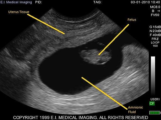
The ultrasound shows many critical pregnancy structures and embryo measurementseight essential features are described below.
How to read ultrasound measurements. I remember lying on that little exam table my husband by my side and splitting my attention between the glowing image on the screen and the ultrasound technicians face. Perform your post-processingeven multi-dimensional analysesoffline on images that retain the same excellent quality as those originally acquired and viewed on your ultrasound system. A nuchal scan or nuchal translucency NT scanprocedure is a sonographic prenatal screening scan to detect chromosomal abnormalities in a fetus though altered extracellular matrix composition and limited lymphatic drainage can also be detected.
This limit varies from person to person and is approximately 20 kilohertz 20000 hertz in healthy young adults. The best portable ultrasound machine presented by EagleView from Wellue Health. Amos Grunebaum MD FACOG is a Professor of Obstetrics and Gynecology and among the worlds leading authorities on fertility and pregnancy.
How to Read an Ultrasound Picture. This unique point-of-view picture generated in real time yields information that goes beyond what is possible with routine imaging methods such as coronary angiography performed in the cath lab or even non-invasive Multislice CT scans. The bioinspired system uses ultrasound measurements of muscle dynamics to develop a personalized and activity-specific assistance profile for users of the exosuit.
Acquiring the IVC view with Ultrasound. An ultrasound is a painless diagnostic test that most women will receive during pregnancy. The candidate should be keen and enthusiastic and have an interest in one or more areas of ultrasound application andor its use in clinical practice.
Ultrasound system operation and optimization including measurements. Designed for healthcare professionals. Ultrasound is widely available easy-to-use and less expensive than other imaging methods.
Introduction and integration of exam protocols. Hands On Ultrasound Training. EagleView wireless portable doppler ultrasound contains both a curvilinear ultrasound probe and a phased array ultrasound probe.
Hand-Eye coordination and beam to screen relationship. CT Scan costs range from 1200 to 3200 - they are usually more expensive than ultrasonography. Ultrasound scanning gives a clear picture of soft tissues that do not show up well on x-ray images.
Intensive 4-5 Day or Weekend Ultrasound Training Courses. Most people will know how to perform an IVC exam but for a more in-depth tutorial on how to obtain the IVC view you can click HERE. Vscan Extend R2 with Dual Probe SonoSim 365.
Ultrasound Training for all Medical Workers including Hospital ER Physicians Paramedics OB-Gyn Sonography and all Ultrasound Training. Point-of-care imaging on your palm. Get our Childbirth Course via email to learn all about labor pain relief natural options and get ready for baby and breastfeeding.
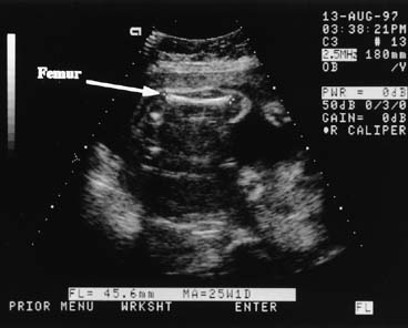
Amos full bio the book about him Lessons in Survival. Biparietal diameter is used to estimate fetal weight and gestational age. Ultrasound the journal of British Medical Ultrasound Society is looking to appoint a suitable candidate as deputy editor.
The A-scan ultrasound takes measurements of your eye. The images obtained measurements can be viewed or stored for use later. I remember my first ultrasound.
Help to optimize your workflow by deciding when and where an echo exam is accessed reviewed or finalized based on your scheduling needs and the expertise of your team. Ultrasound in Medicine and Biology is the official journal of the World Federation for Ultrasound in Medicine and Biology. My excitement ebbed and flowed based on micro-expressions I thought she was makingthough in reality she remained professionally stoic.
Applications of Ultrasound. The journal publishes original contributions that demonstrate a novel application of an existing ultrasound technology in clinical diagnostic interventional and therapeutic applications new and improved clinical techniques the physics engineering and technology of. Place the probe in the subxiphoid position and get the IVC in the long-axis view.
To read an ultrasound picture look for white spots on the image to see solid tissues like bones and dark spots on the image to see fluid-filled tissues like the amniotic fluid in the uterus. Intravascular Ultrasound or IVUS allows us to see a coronary artery from the inside-out. Amos is using his vast experience to launch.
Scan-IQ This exclusive tool introduces new users to essential skills required for ultrasound. Stage 5 The fetal measurements are reviewed in order to look for any abnormalities in the structure. High-frequency sound waves travel into your uterus and they bounce back from the fetus as vibrations.
Biparietal diameter BPD is one of many measurements that are taken during ultrasound procedures in pregnancy. Evaluate the size and collapsibility of the IVC. Handheld ultrasound system with both linear and sector transducers in a single probe.
The cost might vary in different countries. If youre 12 weeks along in the pregnancy you may be able to make out your babys head and if youre 20 weeks along you may even see the spine heart feet and eyes. It is a measurement of the diameter of a developing babys skull from one parietal bone to the other.
Interpretation of IVC Measurements. Ultrasound probe for iPhones and Android phones. A shorter than normal humorous or femur bone or an absence of the fetal nasal bones is sometimes indicative of Down Syndrome.
The combined procedure A. Prior to eight weeks gestation transvaginal ultrasound provides by. All About Amos and a fictionalized account of his fathers life in the novel Through Walters Lens In addition to his current work Dr.
This package includes one Vscan Extend R2 Dual Probe with standard components an additional rechargeable battery one external battery charger a 1-hour session of remote clinical applications training and the SonoSim 365 for GE Healthcare training solution. Since chromosomal abnormalities can result in impaired cardiovascular development a nuchal translucency scan is used as a screening rather than. Ultrasonic measurements of blood flow velocity were based on the Doppler effect with piezoelectric transducers that transmit ultrasound waves with a frequency into the skin.
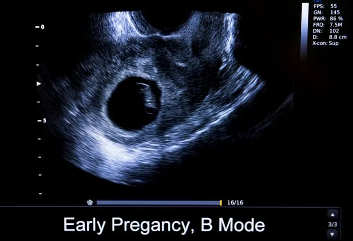
A transvaginal ultrasound an ultrasound in which a probe is inserted into your vagina to get better access to your uterus is generally much more accurate than an abdominal ultrasound in early pregnancy. An ultrasound technician will gently move a transducer or ultrasound wand covered in warm gel around your abdomen to obtain key measurements. Ultrasound imaging does not use any ionizing radiation.
Ultrasound devices operate with frequencies from 20 kHz up to. Get ultrasound images in seconds. Type of Ultrasound.
The B-scan allows the doctor to see the structures in the back of your eye.
Crl Measurements In Pregnancy

Prenatal Ultrasound Scans

20 Week Ultrasound Everything You Want To Know
Diagnostic Obstetric Ultrasound Glowm

Early Pregnancy No Heartbeat Pls Help Read This Ultrasound Report October 2019 Babies Forums What To Expect

Ultrasound Elastography Is A Quick And Non Invasive Way To Spot Kidney Failure Physics World
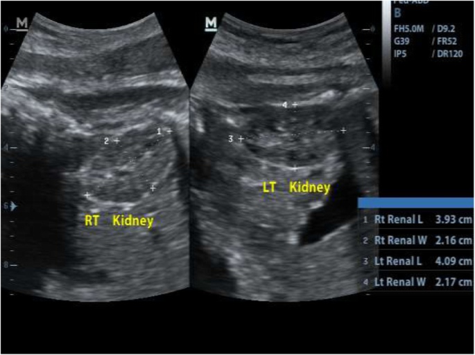
Sonographic Estimation Of Gestational Age From 20 To 40 Weeks By Fetal Kidney Lengths Measurements Among Pregnant Women In Portharcourt Nigeria Bmc Medical Imaging Full Text

Reading Numbers On An Ultrasound September 2019 Birth Club Babycenter Australia


